Daily Archives: October 21, 2010

Life Extension through Nanotechnology

There are two ways in which nanotechnology may be able to extend our lives. One is by helping to eradicate life-threatening diseases such as cancer, and the other is by repairing damage to our bodies at the cellular level – in essence, a nano version of the fountain of youth!

Our average lifespan has been increased over the last 100 years by reducing the impact of life-threatening diseases. For example, vaccines have virtually eliminated smallpox. The application of nanotechnology in healthcare is likely to reduce the number of deaths from conditions such as cancer and heart disease over the next decade or so. There are many research programs working on these techniques;

Let’s look at the type of nano work that is currently being done in the way of eradicating cancer, one of the most serious of diseases on our planet:

An intriguing cancer treatment uses one nanoparticle to deliver a chemotherapy drug and a separate nanoparticle to guide the drug carrier to the cancer tumor. Nanorods circulating through the bloodstream exit where the blood vessels are leaking at the site of cancer tumors. Once the nanorods accumulate at the tumor they are used to concentrate the heat from infrared light, heating up the tumor. This heat increases the level of a stress related protein on the surface of the tumor. The drug carrying nanoparticle (a liposome) is attached to amino acids that bind to this protein, so the increased level of protein at the tumor speeds up the accumulation of the chemotherapy drug-carrying liposome at the tumor. Magnetic nanoparticles that attach to cancer cells in the blood stream may allow the cancer cells to be removed before they establish new tumors.

Read more about nanotechnology and its use in detecting and treating cancer

Another major killer in our time is heart disease. In this area, there are several efforts going on:

Researchers at the University of Santa Barbara have developed a nanoparticle that can deliver drugs to plaque on the wall of arteries. They attach a protein called a peptide to a nanoparticle which then binds with the surface of plaque. Studies have verified that the peptide attaches the nanoparticle to plaque. The researchers plan to use these nanoparticles to deliver imaging particles and drugs to both diagnosis and treat the condition.

Read more about this study of nanotechnology and heart disease

Perhaps the most exciting possibility exists in the potential for repairing our bodies at the cellular level. Techniques in Nanorobotics are being developed that should make the repair of our cells possible. For example, as we age, DNA in our cells is damaged by radiation or chemicals in our bodies. Nanorobots would be able to repair the damaged DNA and allow our cells to function correctly.

This ability to repair DNA and other defective components in our cells goes beyond keeping us healthy: it has the potential to restore our bodies to a more youthful condition. This concept is discussed by Eric Drexler, Ph.D., an established researcher and author whose work focuses on advanced nanotechnologies and directions for current research.

Drexler states: “Aging is fundamentally no different from any other physical disorder; it is no magical effect of calendar dates on a mysterious life-force. Brittle bones, wrinkled skin, low enzyme activities, slow wound healing, poor memory, and the rest all result from damaged molecular machinery, chemical imbalances, and mis-arranged structures. By restoring all the cells and tissues of the body to a youthful structure, repair machines will restore youthful health. ”

Read more about the advances of Nanotechnology from Eric Drexler’s book: Engines of Creation, The Coming Era of Nanotechnology

Maria Konovalenko
SCIENCE FOR LIFE EXTENSION FOUNDATION
http:/mariakonovalenko.wordpress.com/
maria.konovalenko@gmail.com


2 Comments

Filed under Life Extension

First Clinical Trial of Human Embryonic Stem Cell Therapy in the World Begins

Human embryonic stem cell therapy is being tried on a human for the first time in a new clinical trial. This is the first clinical trial of its kind in the world. The trial is designed to test the safety of the treatment, not how well it works. Nonetheless, this is a huge step for regenerative medicine, embryonic stem cell research and science in general!

Working in a handful of medical centers around the country, the biotech firm Geron is treating eight to 10 recent paraplegics. The patients will receive an injection of neurons to the site of the damage, followed by a short treatment of anti-rejection drugs. The first patient is reported as a patient in an Atlanta spinal cord and brain injury rehabilitation hospital. To take part in the study, the patient had to have suffered a spinal or brain injury that resulted in paralysis from the chest down. This patient was injected with cells derived from human embryonic stem cells obtained from a fertility clinic. Researchers are optimistic that this human embryonic stem cell therapy will not only help alleviate the symptoms of the injury, but permanently repair the damage that caused the paralysis from the spinal cord injury.

Embryonic stem cell-derived neural cells have been used by researchers to treat nervous system disorders in animal models. In the case of spinal cord injuries, neural cells derived from animal embryonic stem cells and injected into the spinal cord injury site produced significant recovery of the animal’s ability to move and bear weight.

To apply those observations to humans, Geron had derived oligodendrocyte progenitor cells (GRNOPC1) from Human Embryonic Stem Cells (HESCs). “Initiating the GRNOPC1 clinical trial is a milestone for the field of human embryonic stem cell-based therapies,” said Thomas B. Okarma , Geron’s president and CEO. “When we started our work back in 1999, many predicted that it would be a number of decades before a cell therapy would be approved for human clinical trials. This accomplishment results from extensive research and development and a succession of inventive steps to enable production of cGMP master cell banks, scalable manufacture of differentiated cell product, and preclinical studies in vitro and in animal models of spinal cord injury, leading to concurrence by the FDA to initiate the clinical trial.”

Stem cells have attracted huge scientific and public interest, not only because they bear the promise of miracle cures for age-related diseases, but also because their medical use is so appealing: stem-cell therapies like those that have recently begun could augment the human body’s own regenerative capacity, which declines as we grow older. The appropriate source of cells for these applications is hotly debated, but the technical feasibility of generating replacement tissues and organs is now within realistic projections.

Read more about the human embryonic stem cell therapy clinical trials

Maria Konovalenko
SCIENCE FOR LIFE EXTENSION FOUNDATION
http:/mariakonovalenko.wordpress.com/
maria.konovalenko@gmail.com
2 Comments

Filed under Regenerative medicine

Bioprinting: Laboratory Grown Body Parts Now a Reality

Your liver is failing critically. A transplant would save your life, but there’s a long waiting list and the odds are stacked against you. So instead, doctors extract some of your bone marrow, liver and muscle cells, go back to their laboratory and return in a few weeks with … a freshly grown liver! Does this sound like material from a Hollywood sci-fi movie? Well Not anymore. Australian researchers in Melbourne are now hard at work growing spare parts, proving their stuff in animal – and even human trials!

“It’s a remarkable field, with enormous potential,” says Wayne Morrison, director of the O’Brien Institute at St Vincent’s Hospital in Melbourne and professorial fellow in the department of surgery at the University of Melbourne.

“There is a sense of steady advance,” Morrison says, noting that a key reason for progress is the multidisciplinary nature of tissue generation and regeneration.

“Historically, there has not been much interaction between surgeons, cell biologists, as well as chemical engineers and designers. I’m pleased to say that is changing with both the practical people and the theory experts learning a great deal from each other.”

Morrison is a plastic and reconstructive surgeon. Part of his work involves transplanting blood vessels from a healthy part of a patient’s body to a diseased or injured part to aid recovery. He noticed the new blood vessel would start to promote tissue growth, although the process wasn’t easy to observe.

“We came up with the idea of enclosing the [growing tissue] in a plastic chamber that was surgically inserted into the patient’s body,” he says, explaining that the idea was to see what was happening.

“We were surprised that the chamber quickly filled up with healthy tissue around the vessel. The chamber was acting like a scaffold, a magnet for cells. So in trying to do one thing we found something that was even more important.”

Further experiments revealed that the location of a chamber determined the types of tissue that were generated. Morrison says it’s not clear how this happens but he suggests it may be linked to the proximity of other types of cells, which cue the formation of the new tissue.

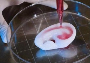
When it comes to growing new tissues for transplants, the truly sci-fi advance is the bio-printer a device that “prints” three-dimensional tissue on to a template. A Melbourne company Invetech developed the technology for Organovo, a biotech firm based in San Diego, California.

“It was a matter of applying Invetech’s expertise in engineering and automation with Organovo’s background in regenerative medicine,” says Invetech’s managing director Fred Davis. “This collaboration was important not only on the robotics side, but also in areas like the software that controls the living cell printing process, where Organovo biologists worked with Invetech software engineers to provide easy-to-use shape programming.”

The bio-printer works by dispensing cells taken from the patient’s body, layer by layer, to create a 3-D tissue or organ. A gel is used to fill in gaps and provide physical support while the cells organize themselves into patterns and bind together. The gel is removed in the lab during a maturation process and the fully formed tissue or organ is then ready for the surgeon to transplant into the patient.

Invetech delivered the first bio-printer to Organovo late last year and Davis predicts production models will roll out soon.

It would seem as though science is really making head way into the future. While creating living tissue is important, the larger aim for science and medicine is creation of functioning human organs. Scientists have very high hopes that one day on the horizon not only organs can be grown in a lab but also nerves and tendons lost in such cases as spinal cord injuries and paralysis. Organ replacement will eventually enable humans to have indefinite lifespans through complete rejuvenation to a youthful condition.

Read more about tissue regeneration and organ growth research

Here’s a fascinating video on the 3D Bio-Printer

Maria Konovalenko
SCIENCE FOR LIFE EXTENSION FOUNDATION
http:/mariakonovalenko.wordpress.com/
maria.konovalenko@gmail.com

9 Comments

Filed under Regenerative medicine